
Collie Eye Anomaly (CEA)
With Eye diseases, whether there is a DNA test or not, the ANKC Australian Canine Eye Scheme (ACES) is a National Assessment System for ANKC registered dogs. It offers qualified certification for a range of congenital and inherited eye conditions, with the assessments being carried out by registered Veterinary Eye Specialists. Please go to the Eye Health and Screening page of our website for futher information.
Collie Eye Anomaly (CEA) is one of the diseases that has DNA testing available and if a dog is showing as an 'affected', then examination by a register Veterinary Eye Specialist should be done to confirm the status of the disease in your dog.
 |
 |
Inheritance: Autosomal Recessive (with incomplete penetrance, this is when some individuals who carry the CEA disease gene, develop CEA, while others do not)
Symptoms: Severity of the disease varies. The mild form, choroidal hyperplasia (CH), does not cause visual defects and blindness. Severely affected dogs have colobomas which can result in reintal detachments and bleeds causing blindness. This can occur in pups or suddenly and usually by the time a dog is 2 years old.
Description:
CEA is an inherited disease that causes defects in the formation of the eye resulting in visual impairment. It affects a number of breeds in the Collie family.
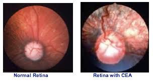
Several aspects of the disease are recognized, however the most important is a lesion (defect) on the back of the eye called choroidal hypoplasia (CH) which can only be seen opthalmoscopically. The choroid is a layer of tissue in the eye responsible for supplying blood and nutrients to the retina. In dogs with CEA, the choroid does not develop properly and is thinner than normal. The severity of this lesion can vary from dog to dog.
 All dogs affected by CEA have choroidal hypoplasia, by definition. More severely affected dogs may have pits (colobomas) affecting the retina and nearby tissues and in the most severely affected eyes, retinal hemorrhaging and detachments can occur, resulting in blindness. The severity of the disease varies with mildly affected dogs producing severely affected/blind puppies and vica versa. The disease is not progressive like prcd-PRA and most affected dogs may only have mildly impaired vision.
CEA can be detected by a physical eye examination performed by a veterinary opthalmologist between 5-12 wo (preferably by 8 wo). After that age-related pigmentation of the retina often masks the characteristic, disease-related changes. These are the dogs referred to as "go-normals". This results in a false negative result. DNA testing will confirm whether a dog is affected or a carrier.
The CEA/CH mutation was discovered by Dr Aclund at the University of Cornell with the DNA test becoming available in 2005.
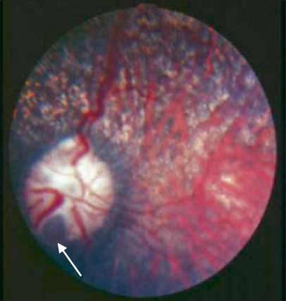
Above an eye with Choroidal Hypoplasia/Colobomas
Varying degrees of CEA and definitions are listed below:
-
Choroidal Hypoplasia, Chorioretinal Change, Choroidal Dysplasia: These refer to abnormalities in the coloring or pigmentation of the choroid or central layer of the eye's lining. This is the most common abnormality found in CEA and all dogs with CEA have CH. However it is the least harmful and mildest form of CEA. Most dogs with this form function normally with no ill-effects or loss of vision.
-
Coloboma, Ectasia, Staphyloma: While not completely synonymous, these terms all refer to a cupping or bulging in the eyeball usually in the area of the optic nerve. Colobomas are the most common more serious complication of CEA. Colobomas can be described as a pit in the eye or a blister at the back of the eye that you can see with an ophthalmoscope. Colobomas vary and can be small or large and occur in approximately 25% of dogs with CEA.
-
A third set of complications which occur exclusively in dogs with coloboma include retinal detachment or hemorrhaging in the eye. About 5-10% of dogs with CEA have these severe complications, which can lead to blindness. These percentages are based on experience with the Collie Breeds.
-
Vascular Disease, Tortuous Blood Vessels: These terms describe defects in the vessels of the eye, which are responsible for its blood supply or "nourishment." These may be malformed, undersized, or even lacking.
-
Retinal Detachment: Loosening or separation of the innermost, or retina, layer from the wall of the eye. This may involve a tiny area or the entire retina. It can be either one or both eyes. The complete detachment of the retina results in blindness in that eye.